From core diagnostic imaging to specialized MRI technology, DFW MRI offers advanced services designed to support better comfort, faster workflows, and more detailed imaging across our DFW locations.
Uses magnetic fields to create detailed images of soft tissues, joints, organs, and internal structures.
Read More →Combines multiple X-ray images to provide detailed cross-sectional information on bones, tissues, and organs.
Read More →Provides fast, reliable imaging used to evaluate fractures, injuries, arthritis, and other bone-related concerns.
Read More →Diffusion Tensor Imaging provides advanced visualization of white matter pathways to support neurological evaluation.
Advanced MRI reconstruction technology designed to improve image clarity while helping support efficient scan performance.
Specialized MRI coils help optimize signal capture for targeted anatomy, supporting detailed and high-quality imaging.
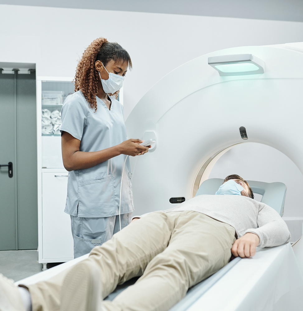
An MRI (or magnetic resonance imaging) scan is a radiology technique which uses magnetism, radio waves, and a computer to produce images of body structures. The MRI scanner is a tube surrounded by a giant circular magnet. The patient is placed on a moveable bed which is inserted into the magnet. The magnet creates a strong magnetic field which aligns the protons of hydrogen atoms, which are then exposed to a beam of radio waves. This spins the various protons of the body, and they produce a faint signal which is detected by the receiver portion of the MRI scanner. The receiver information is processed by a computer, and an image is then produced.
The image and resolution produced by MRI is quite detailed and can detect tiny changes of structures within the body. For some procedures, contrast agents such as gadolinium are used to increase the accuracy of the images.